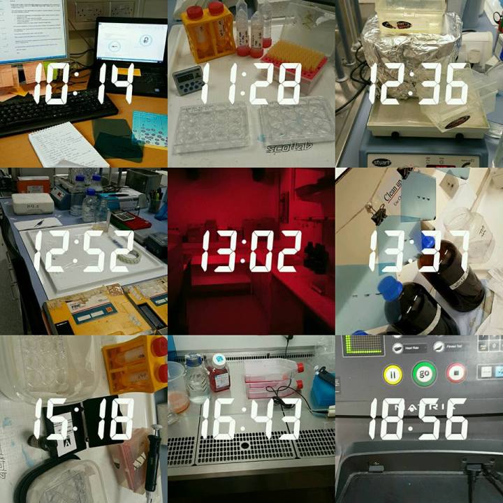
Hello and welcome to yet another insight into my daily life as a PhD student in a stem cell lab!
After a slow January with cells and experiments, February is here and the incubator is overflowing with cells – mainly for my experiments actually so I thought it was time to update you with another day in the life of a PhD student post!
.
7:42 am – Alarm goes! Time to wake up and get to it! Today I wasn’t heading straight into the lab. I had some ‘admin’ to sort out at home first and actually having breakfast – which is basically unheard of for me!
.
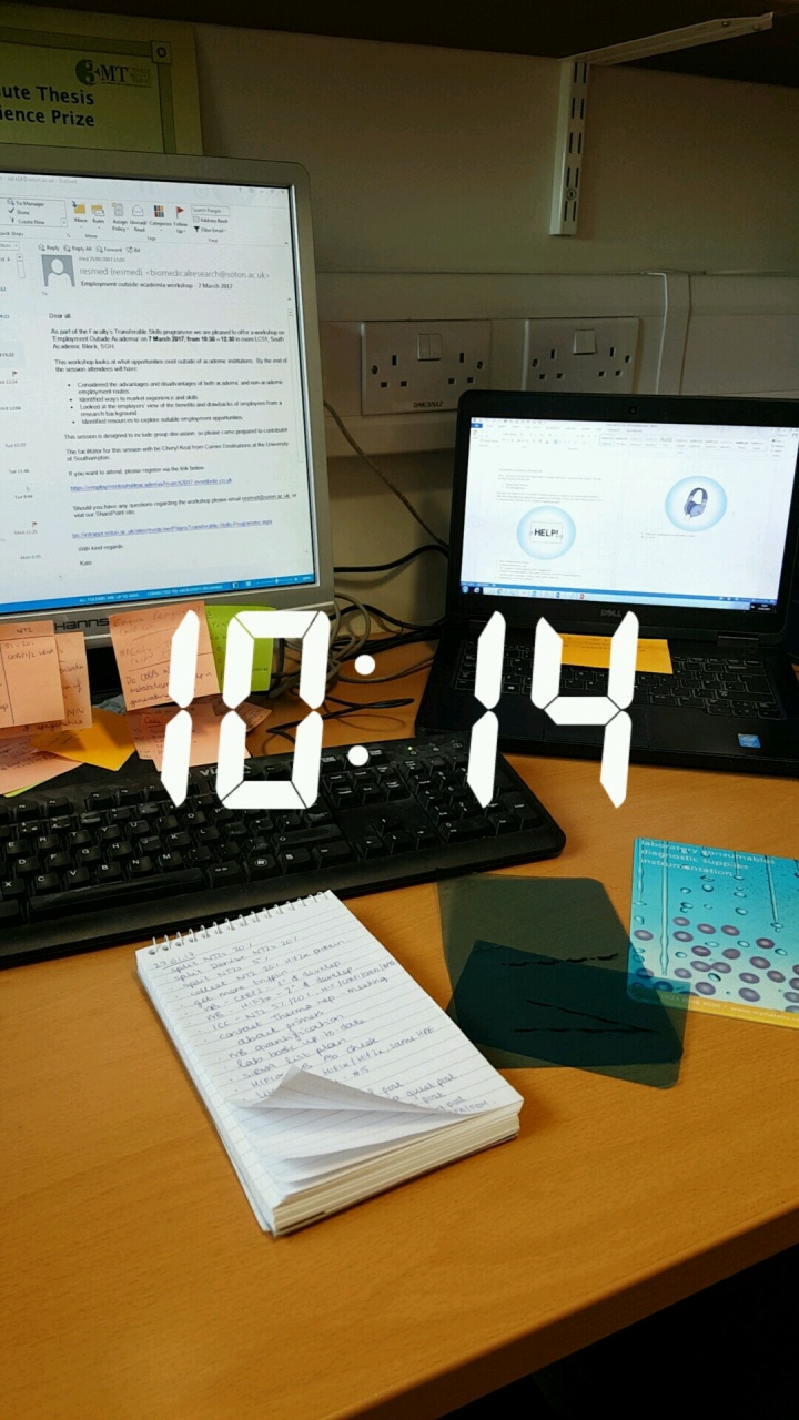
10:14 am – Finally made it to work! The joys of working in academia – means I can roll into work at this time if I need to! So, to start off my working day, it was the usual checking emails, labelling any experiments I had finished the previous day etc. I’m also trying to write a guest blog post so I had that open ready to write during all the incubation steps I had during my experiments for the day – watch this space for that guest blog post being published soon!
But it was time to actually face the lab and doing experiments. So today I embark on the second day of our Western blotting protocol. I’ve shown you the first day way back in Chapter 1 of this series but now you can get a flavour of what the next steps after that were. So my membranes have been in primary antibody overnight so we wash our membranes first thing in the morning to remove any unbound antibody and then add a secondary antibody. These antibodies recognise the primary antibody so increase the intensity of our protein band.
.
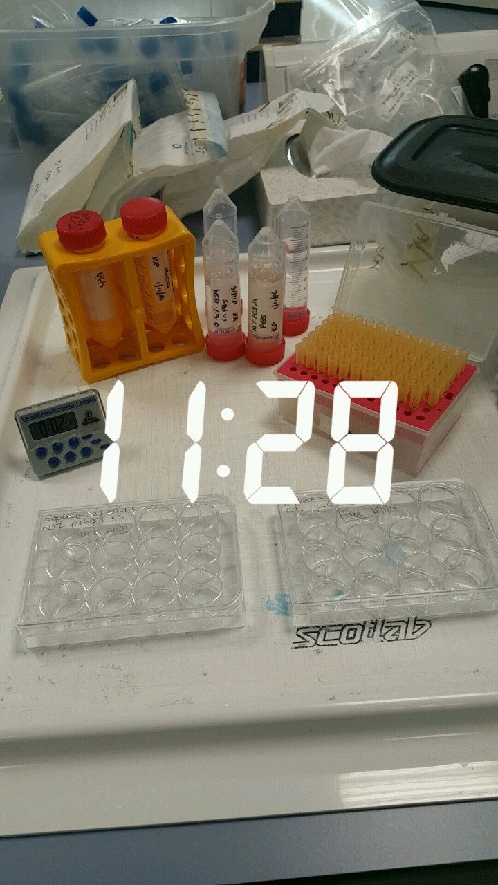
11:28am – I get an hour long break now from the Western blot! But I’m starting my second experiment of the day. I want to do an immunostaining on my cancer stem cells to see whether they are expressing my protein of interest and it’s the method of how I get the blue and green images I have shown in my previous ‘Cellfie’ of the month blog posts. And if you want to learn more about the cancer stem cells I’m researching check out February’s ‘Cellfie’ of the month. I won’t go into the details of what I have done for this technique as I’ve gone through it before – but if you’re new to my blog or missed it just follow this here but it’s basically another technique I use to see where my protein of interest is expressed in my cells using antibodies. It uses the same principle as Western blotting!
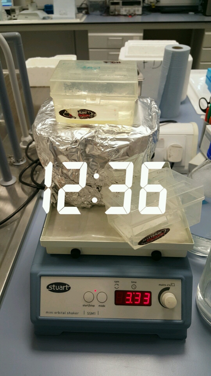
12:36pm – I seem to be doing too many experiments for the one shaker we have in the lab. I’ll just hope noone else needs to use it But the primary antibodies have been added to my immunocytochemistry plates and they are now binding to the proteins in my cells in the dark. And the secondary antibody has been washed off the membrane now and it’s time to develop the blot!
.
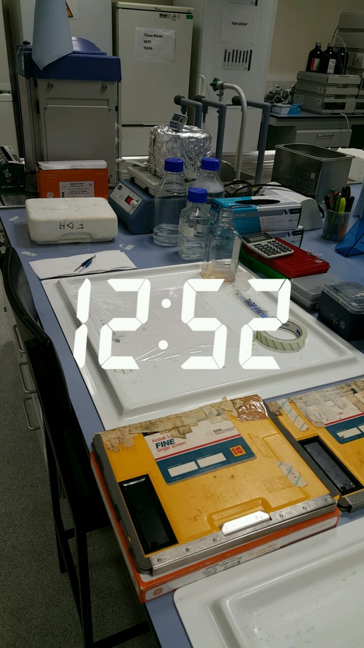
12:52pm – The set up for Western blot development in our lab is old school! We use X-ray films and the set up takes up a lot of space! So basically – on our membranes are a series of protein bands that are all separated by size by they are invisible at the moment! The development stage is where we make these bands visible but hopefully we will only get the one or two bands appear that are specific to the protein we are interested in. Fingers crossed that the protein band that contains our protein of interest has been bound by the primary antibody and now all of those primary antibodies have been bound by a secondary antibody.
On the end of our secondary antibody though is a tag or a label and when we add our detector fluid this is going to switch on that tag which will show us where on our membrane the antibodies have bound – which should be where our protein is!
.
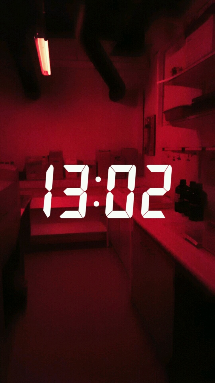
1:02pm – Detector fluid added and the clock has started! The tag on the end of the secondary antibody will only be switched on for a short while so we need to use the X-ray film to see where they are bound before it turns off again!
And we do this in the dark room! And it is where I spend about 99.99% of my lab time – or at least it feels like I spend that amount of time in here! Now this might remind you of films where characters are sat under some red light developing photographs? That is basically what I try to achieve every time I go in here except I’m developing a picture of my protein bands using the Xray film! There are lots of more high tech ways to develop Western blots now – maybe some of you reading this do it a different way, if you do please comment below so others can learn about the way you do it 🙂 – but we stick to this method as it is the best for the stem cells we are researching!
.
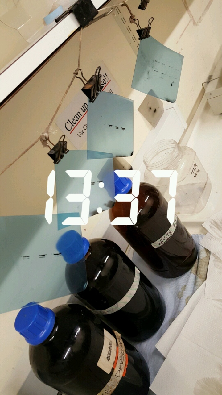
1:37pm – After a host of different exposure times and quite possible different membranes if I’m looking at different proteins – this is what I get! A small piece of blue xray film which has black bands on it! These are those invisible protein bands I’ve been telling you about but you can now see them!
For this particular experiment I was looking at four different samples, hence you can see four bands horizontally – one for each sample. The same amount of protein has been added to the gel for each sample so some bands appear darker than others because there is more of that protein in that sample compared to the other ones!
.
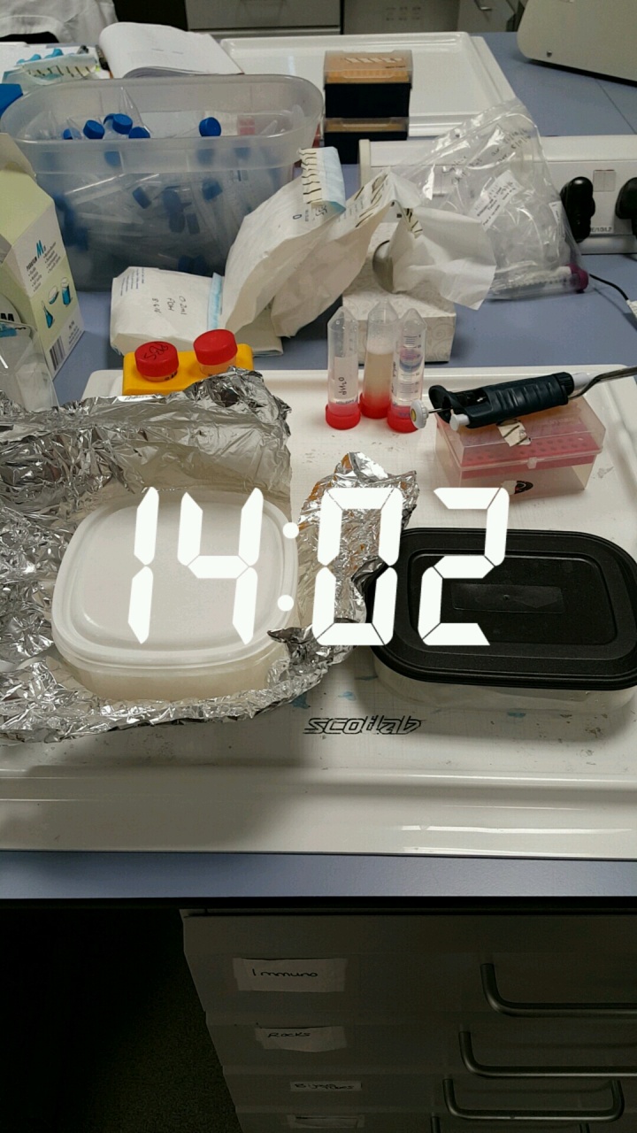
2:02pm – Back in the main lab now and time to add the secondary antibody to my immunostaining plates. The plates are inside the boxes – I promise I’m not eating my lunch in the lab!
But also, we need to check that the differences in the band intensity from my Western blot are due to a biological reason and not just because I loaded the wrong amount. So I’ve now added my membranes to an antibody which will bind to what we call a ‘housekeeping gene’. What this means is that it recognises a protein that doesn’t change expression between cells no matter what conditions we change! So if the bands from this protein are the same, then we know the differences in the bands we saw before are due to a biolgical reason!
.
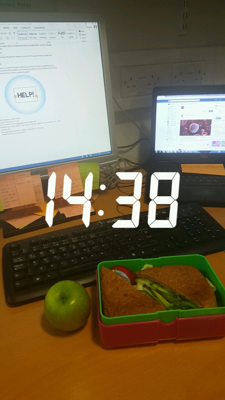
2:38pm – I told you! No lunch in the lab, instead I have a break in my Western blot and my immunostaining now which means I can actually get some food! Thank goodness as my stomach was starting to rumble quite loudly by this point!
.
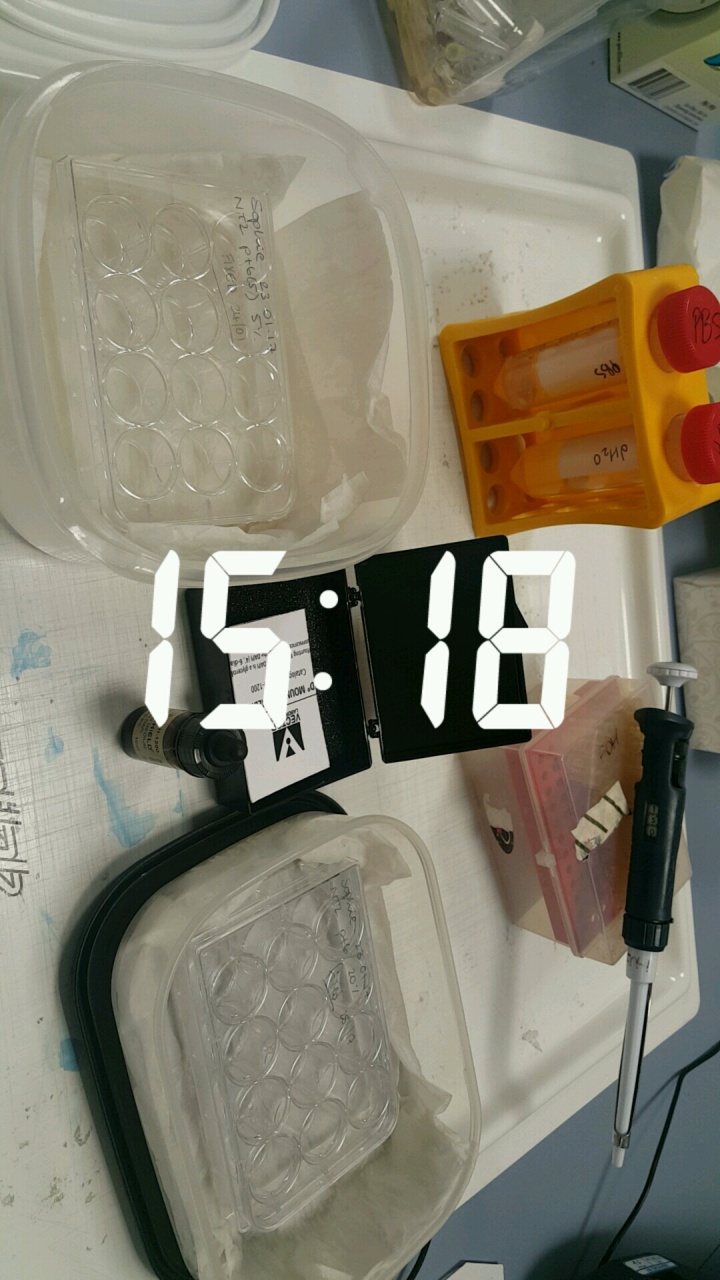
3:18pm – Secondary antibody step is now complete for the immunostaining. Now we need to stain the nuclei of our cells using the DAPI stain I explained in my first ever Cellfie of the month post! Once that is done, the plates are stored in the fridge until I have some time to take some images!
.
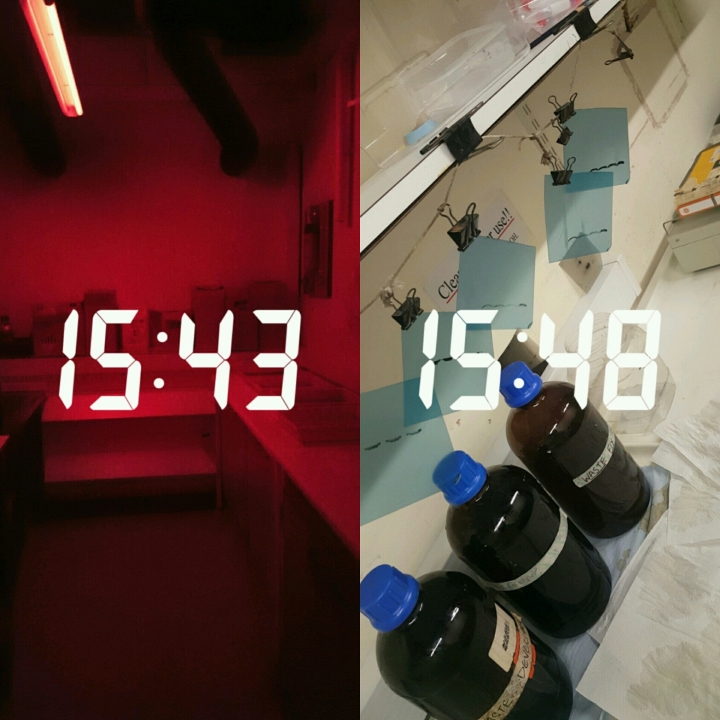
3:43pm – Back to the dark room I go! Time to see if our ‘housekeeping gene’ bands look about the same level. 5 minutes later – I have more pieces of film to add to my ever growing collection! And the bands look good! We have a biological effect 🙂
.
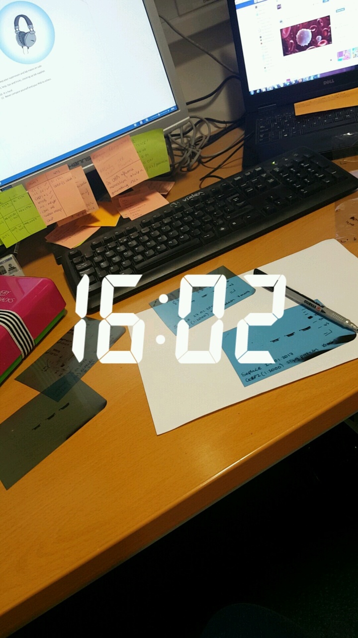
4:02pm – Time to label my pieces of Xray film up so I know what samples these bands are from and what protein I was looking at so I can quantify the expression between my samples later! I also spent a bit of time writing a section of this guest blog post I have been drafting.
.
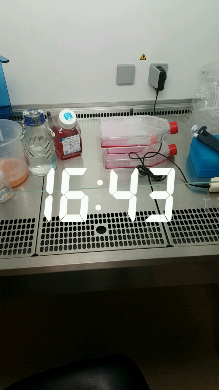
4:43pm – Feeding time in the lab again! So my stem cells have been fed in a different lab but now I’m looking after my cancer stem cells. If we kept feeding and feeding our cells they would keep growing and growing and eventually they would run out of space in the tissue culture flasks or plates that they are in and more than likely die. So in order to keep them growing, we split them every few days. This basically means that I unstick them from the bottom of their flasks and only move half of them into a new flask which means there are less cells in the flask but they have more space now to keep growing!
.
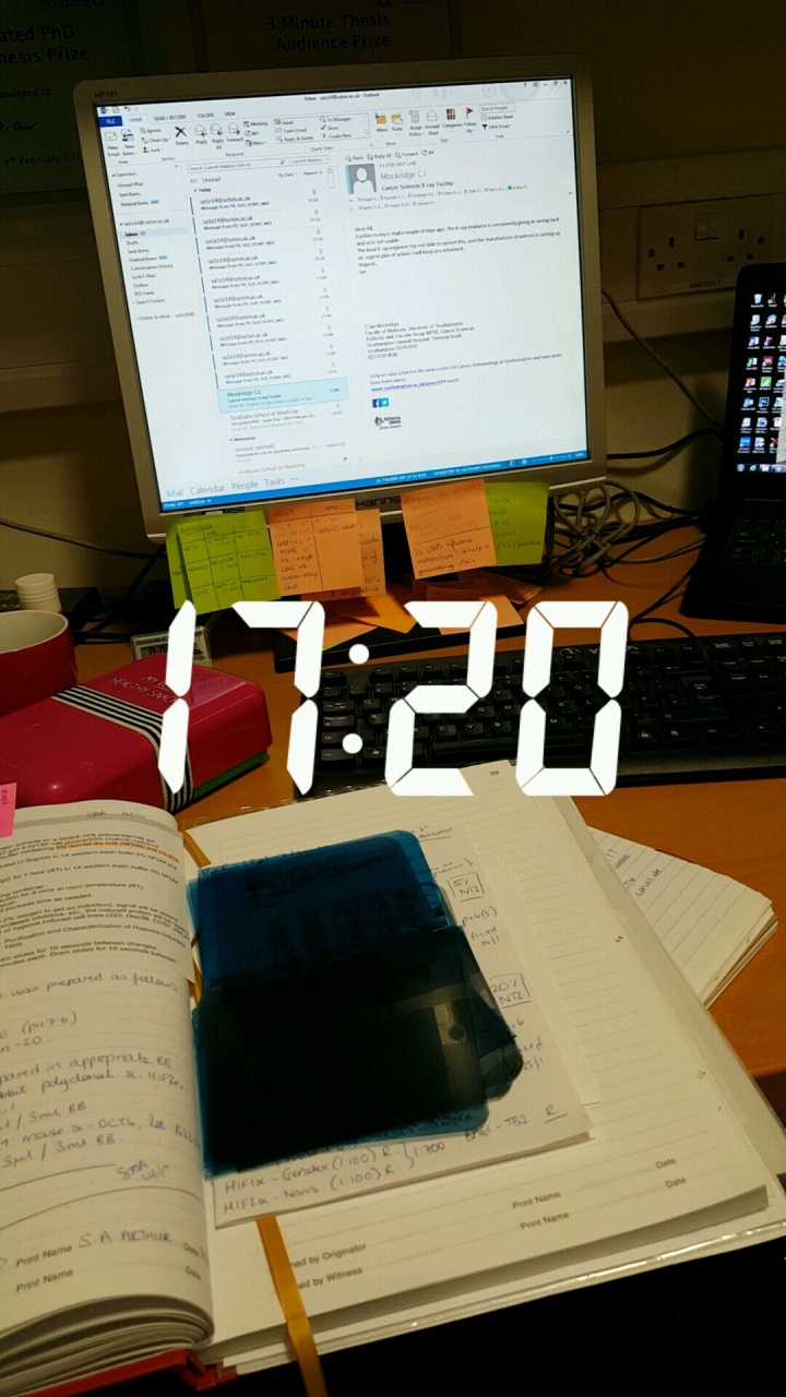
5:20pm – This time is for my end of day admin! I have done the quantification of my Western blot which has shown me some exciting results 🙂 which I would love to share but you’ll have to wait for my publication to see 🙂 And then everything I’ve done in the lab today gets written up into my lab book, followed by a quick check of my emails before turning the computer off and heading home!
.
6:56pm – I wasn’t home for long though! In the door, quick change and back out the door as I need to get a gym session in! I’ve tried to be a lot better this year with the health and exercise malarkey! So far it seems to be going quite well. I’ve also found that I actually quite enjoy it when I’m there – the problem is getting me there! I’ve also found it’s a good stress reliever! As a very busy PhD student, it is a good way for me to release some excess energy before heading back home and can actually chill for the night!
.
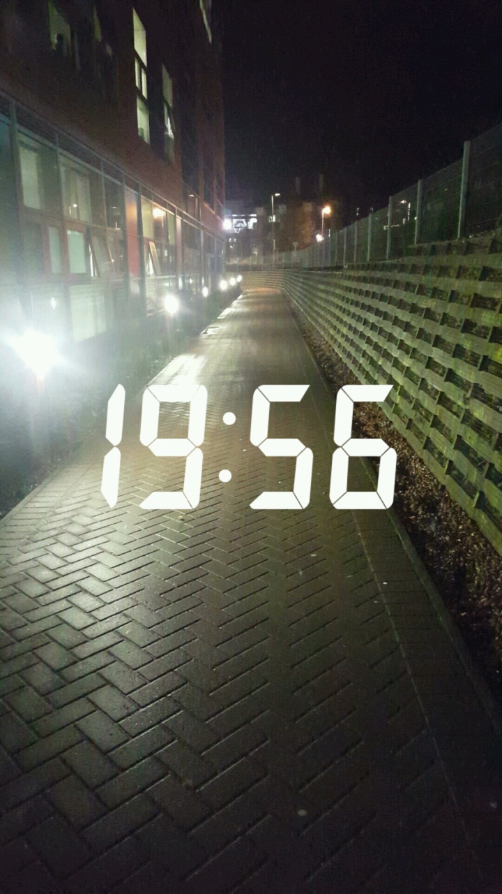
7:56pm – Workout done! Walk home to burn those extra few calories! Time to eat! And probably do a bit more work on my guest blog post, or maybe I’ll just chill and watch some TV.
.
My days are definitely getting busier now and they are definitely getting longer with all my lab work and other scicomm commitments I’m starting! But hopefully I will be organised and manage it all! But I would much prefer to be busy than not have anything to do for days on end.
.
What is a typical day like for you and your science research? Please share your stories so anyone who reads this blog can see how varied a scientist’s day can be! It is not lab 9am-5pm Monday to Friday.
I hope you enjoyed another insight into my life as a PhD student. Please feel free to ask my any questions you might have about what I’ve written about today or my work in general if you like.
